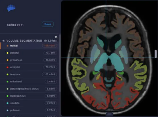
Brain volumetry MDBRAIN
Precise, quantitative statements on atrophic or enlarged structures.
Fully automated.Rapid determination of brain volume.
Precise, quantitative statements on atrophic or enlarged structures. Fully automated or with just one click
Rapid determination of brain volume with differentiation of pathognomonic relevant regions, such as the hippocampus.
For each evaluation, the deviation from the normal value is shown. For this purpose, the results are compared with a comprehensive and heterogeneous database based on age, gender and intracranial volume (approx. 8,000 data sets).
Supports you with the diagnostics of
- Dementia:
- Volumetry of frontal lobes, parietal lobes & temporal lobes (with hippocampus).
- Multiple sclerosis:
- Volumetry of the whole brain & thalamus
- MSA, PSP, CBD:
- Volumetry of nucleus caudatus, putamen, pallidum, cerebellar cortex and mesencephalon.
Capturing atrophy patterns at a glance.
Plotting of objective volumetry results for pathognomically relevant brain regions.
From the resulting patterns, conclusions can quickly be drawn about the underlying disease. The comparison with the last preliminary examination also enables an assessment of the dynamics of a neurodegenerative process.
Detect neurodegenerative processes at an early stage.
Plot of the percentile values resulting from the normal value adjustment over time against the age of a patient.
By including existing preliminary examinations, neurodegenerative processes can thus already be recognised as a deviation from the physiological ageing process before they become reliably pathological as a single value.


 Edificio Torre Europa,


